
Phone
+86 18630938527
The product of time:2022-07-13 14:22:30
Description:
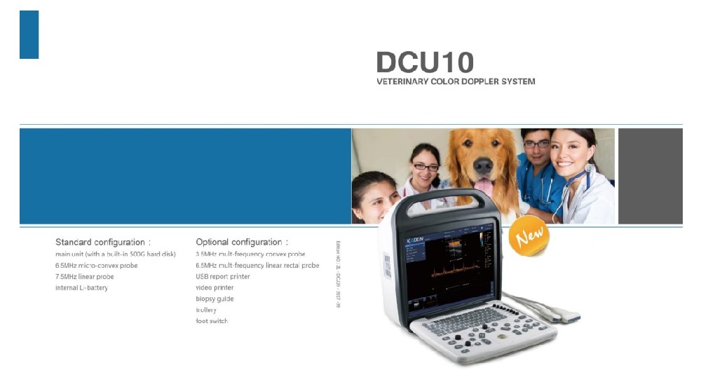
DCU10VETfull digital color Doppler ultrasound diagnostic instrumentDCU10 VET Technical specifications1. Instructions for useApplication range of instrument: DCU10have the leading image process technical and practicalclinic solution method, apply to ultrasound diagnosis...
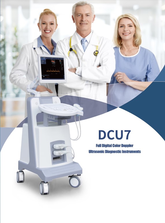
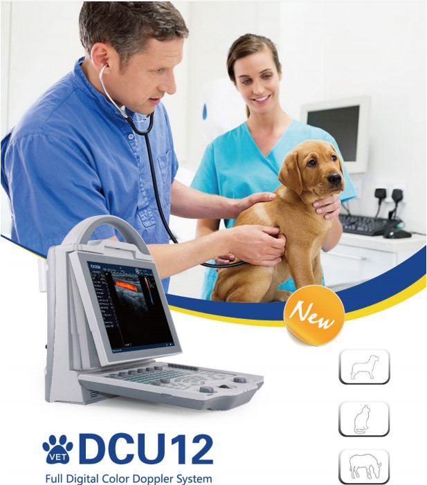
DCU10VET full digital color Doppler ultrasound diagnostic instrument
DCU10 VET Technical specifications
Application range of instrument: DCU10 have the leading image process technical and practical clinic solution method, apply to ultrasound diagnosis of Bovine, Equine, Camel, Canine, Feline, Goat, Ovine and Swine.
For examine of abdomen, heart, gynecology, obstetrics, urology, small organs, blood vessels, etc.
2.1 Machine size, weight
Size: 400(L)*394(H)*172(W)
Weight: only unit 7.5kg, with one probe 8.2kg, with two probes 8.9kg.
Channel: 64 channels;
Probe elements: 128;
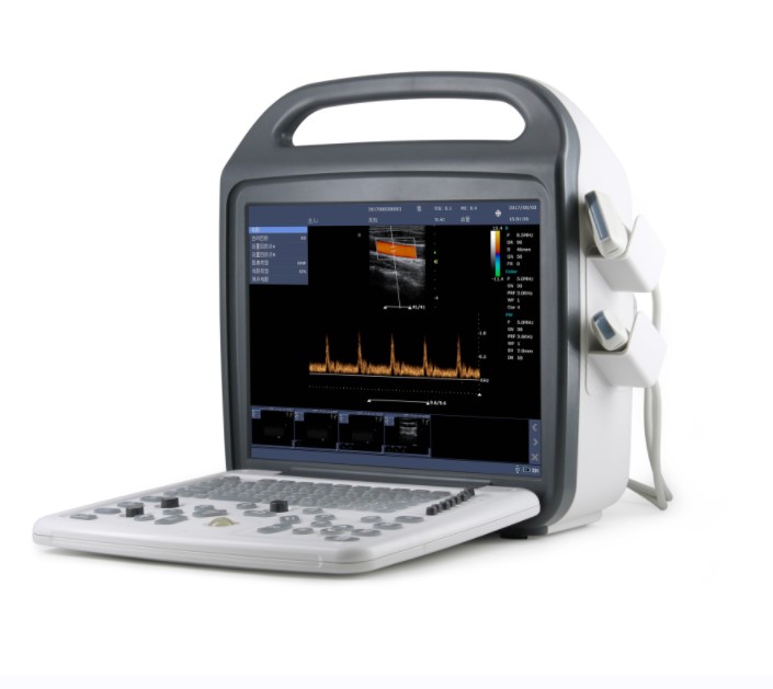
High resolution 15 "LCD display
1. Pulse inverse harmonic composite imaging
2. Multi-beam synthesis
3. Space compound
4. Image enhancement noise reduction
1. B mode
2. M mode
3. Color (Color Doppler ) mode
4. PDI (Energy Doppler)mode
5. PW (Pulse Doppler)mode
2.6 Image display mode
B、2B、B+M、4B、M、B+Color、B+PDI、B+PW、B+Color+PW、B+PDI+PW、★B+C/PDI Double real time
1. Convex probe
2. Linear probe
3. Cavity probe
4. Micro-convex probe
Probe frequency: 2.5-10.0MHz
Probe socket: 2 PC
B/M: Third gear fundamental frequency + second gear harmonic frequency;
C:2 gear
PW:2 gear
1. 2D mode, B Maximum: ≥5000 frames, Color, PDI Maximum: ≥2500 frames;
2. Timeline mode (M, PW), Maximum: ≥ 190s
Real-time scanning(B、B+C、2B、4B)Status: stepless enlargement
1. Support JPG, BMP, FRM image formats and CIN、AVI movie format
2. Support local storage;
3. ★Support DICOM, meet the DICOM3.0 standard;
4. ★Built-in workstations: Support large-capacity hard disk (≥500GB),support patient data retrieval and browsing.
1. Chinese / English operating system and language environment, support other languages according to user requirement;
2. Full screen annotation input, Chinese/ English; Chinese input methods ≥2,including Wubi input method
2.13 Measurement calculation software package
Abdomen, gynecology, obstetrics, small organs, heart, blood vessels, etc.
Built-in high-capacity lithium battery, continue to work more than 1.5 hours, power information displayed on the screen.
3.1 B mode
1. Gray scale mapping:≥15
2. Noise suppression:≥8
3. Frame correlation:≥8
4. Edge enhancement:≥8
5. Image enhancement:≥5
6. Space compound: Switch adjustable
7. Scan density: High, middle, low
8. Image flip: Up, Down, Left, Right
9. Maximum scanning depth:≥320mm
3.2 M mode
1. Scan speed(SweepSleep):≥5, adjustable
2. Line average(LineAverage):≥8
1. SV size / location: SV size1.0–8.0mm , adjustable
2. PRF:≥16 ,0.7kHz-9.3KHz ,
3. Scan speed(SweepSleep):≥5, adjustable
4. Correction angle(CorrectionAngle):-85°~85°,step length5°
5. Spectrum flip:Switch adjustable
6. Wall Filter:≥4, adjustable
7. Doppler sound:≥20, adjustable
1. PRF:≥15 ,0.6KHz –11.7KHz
2. Color map(color map)≥4 kinds;
3. Color related:≥8
4. post-treatment:≥4
1. B/C mode routine measurement: Distance, area, perimeter, volume, angle, area ratio, distance ratio
2. M mode routine measurement: time, slope, heart rate, distance
3. Doppler mode routine measurements: heart rate, velocity, velocity ratio, resistance index, pulse index, manual / automatic envelope, acceleration, time
4. Obstetrics B, PW mode application measurements: EDD and GA for bovine, equine, ovine, canine, feline, goat,swine and llama and OB report generation..
5. Heart B、M mode application measurements
6. Blood vessels PW mode application measurements
7. Small organ B mode application measurements
8. Abdomen B mode application measurements
1. Main unit
2. High-resolution 15-inch LCD monitor
3. 6.5MHz micro-convex probe:1 pc
4. 7.5MHz linear probe: 1 pc
5. Inverse harmonic imaging technique
6. PDI(Energy Doppler mode)
7. Spatial composite imaging
7. Built-in workstation
If you have any questions, please contact us!
CONTACT US