
Phone
+86 18630938527
The product of time:2022-07-14 14:31:31
Description:
DCU12 Full Digital Color Doppler Ultrasound scanner(Vet)1.Instrument name: DCU12 full digital color Doppler ultrasound diagnostic instrument Application range of instrument: DCU12 have the leading image process technical and practical clinic solution method, apply to ultrasound diagnosi...
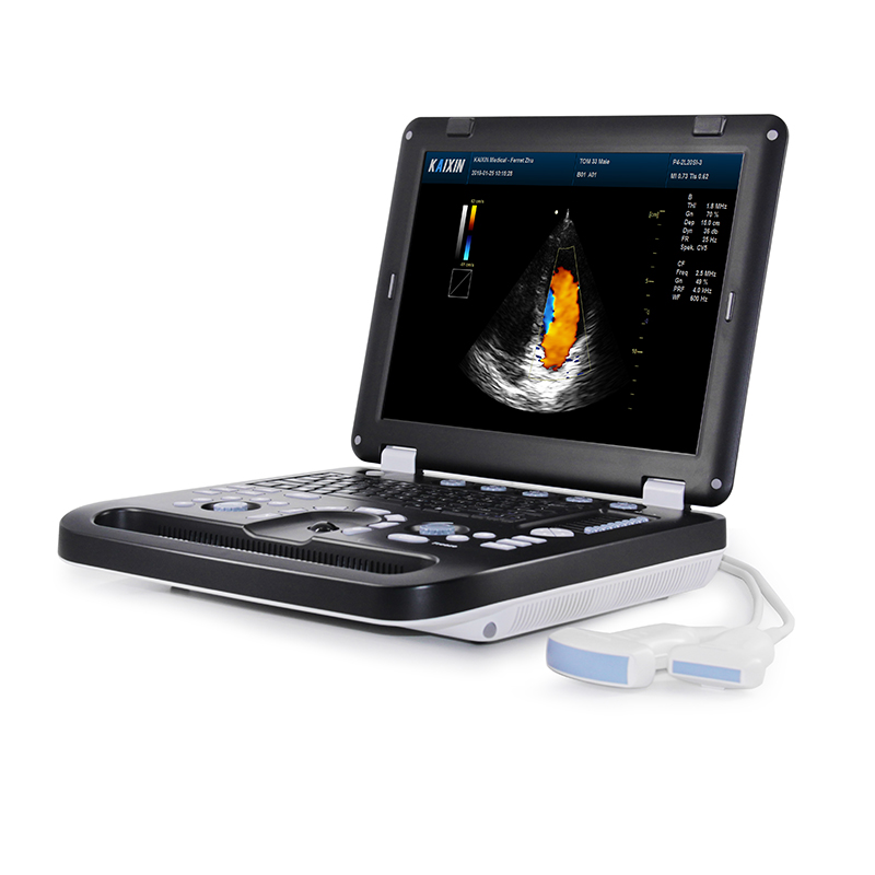
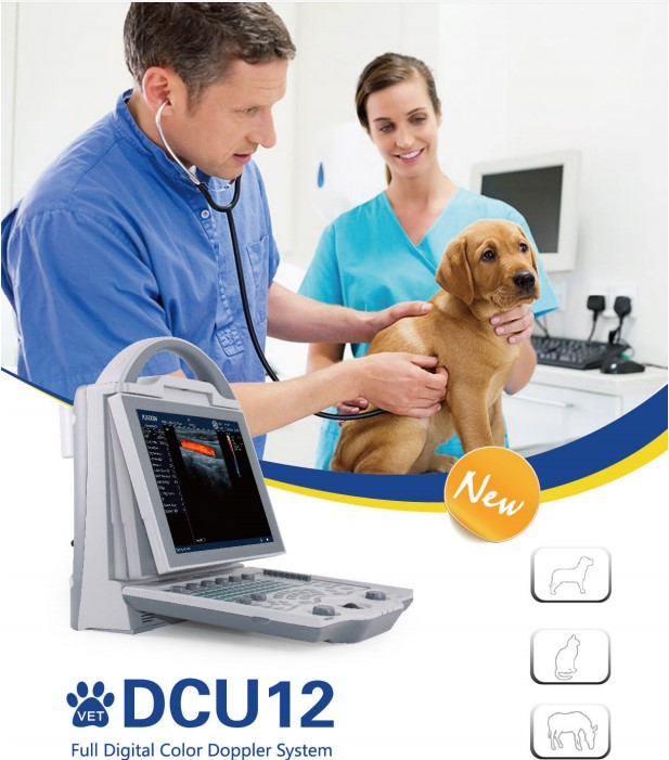
DCU12 Full Digital Color Doppler Ultrasound scanner(Vet)
1.Instrument name:
DCU12 full digital color Doppler ultrasound diagnostic instrument
Application range of instrument: DCU12 have the leading image process technical and practical clinic solution method, apply to ultrasound diagnosis of Bovine, Canine, Equine, Feline, Goat, Camel, Ovine and Swine.
2.Primary system performance overview
1. Adopt A8 Embedded flat form, entirely defenses virus invasion, with highly reliability.
2. Operation system: Linux operation system, Chinese/English menu can switch arbitrarily.
3. Full digital multi-beam formation technologies, high accuracy postpone point wise adopt dynamic focusing, broadband frequency imaging, adaptive imaging optimization technique, adaptive vessel imaging, adaptive Doppler imaging etc.
4. Broadband frequency full digital sound former: dynamic focusing.
5. Two-dimensional gray-scale imaging unit.
6. M mode imaging unit.
7. Dynamic range can visual and adapt.
8. Two-dimensional gray-scale imaging unit, gray-scale≥256, with excellent subtle and contrast resolution and full uniformity.
3. Technique parameters
l Standard parameters
1. Gray scale: 256
2. Color scale: 256
3. Monitor: 10.4” flicker free high resolution medical color LCD.
4. Adapter rating: 100-240V~1.2-6.0A Frequency: 50-60Hz
5. Output of adapter: DC12.8V 3.0A
6. Power consumption: ≤100VA
7. Main unit size: approx. 256*150*326(mm, L*M*H)
8. Weight of main unit: approx. 4.5kg(excluding accessories)
l Multimedia and external device
1. Video recorder;
2. I-Station integral working station, realizing store image, general report and could print function.
3. U-disk (support document management, software upgrading, one-key storage function), DICOM port, convenient for doctor to manage data, support long-distance transmission.
4. Dual-mode TV output: PAL/NTSC
l Probe socket
Two activate probe socket, with automatically identification technique, support optional multi probe.
l Language
Menu interface operation, interface language: can switch Chinese/English
l Probe parameter
Number of elements: 80
Linear rectal probe 6.5MHz; Range of frequency: 4.5/ 5.5/ 6.5/ 7.5MHz
Convex array probe 3.5MHz; Frequency range: 2.0\2.5\3.5\5.0MHz
High frequency broadband probe 7.5MHz; Frequency range:6.0\6.5\7.5\9.0MHz
Dimpling broadband probe6.5MHz; Frequency range:5.0/6.5/7.5/9.0MHz
●Operation mode
1. B、B/B、4B mode
2. M、B/M mode
3. Color flow mode(CFM)
4. Power Doppler mode(PDI)
5. Pulsed-wave Doppler(PW)
6. Tissue harmonic imaging(THI)
●System preset: basic preset,formula preset, note preset, store preset, code preset, reset to default.
4. Ultrasonic imaging technology
1. High precision digitizing continued wave former
Elaborate Ultrasonic beam control to eliminate side lobe noise, increase resolution and contrast resolution greatly, and display the whole structure exquisitely.
2. Dynamic frequency blend imaging technology
Automatic control the sending and receiving frequency of the far field and near filed to combine the powerful penetrating with high definition image perfectly.
3. High precision delay point-by-point dynamic receiving focus
To demonstrate High precision delay point-by-point dynamic focus to the whole image, showing the true and exquisite organize information.
4. Ultra-broadband imaging technology
to choice the wanted central frequency according to different people
5. Automatic optimizing process image technology
Automatic optimize digital parameter according the the current receiving signal to display the perfect ultrasonic image.
6. Automatic vessel imaging technology
Automatic adopt the proper filtering program according the current receiving Doppler deplore echo signal to control stray wave effectively. The perfect combination of resolution with sensitiveness makes sure the blood flow image more clear.
7. Automatic Doppler imaging technology
To strength the weak Doppler signal, and strength the frequency spectrum signal by the complicated digital process technology to improve the Doppler sensitive and display effect.
8. THI structure harmonic imaging technology
To send the ultrasonic in low frequency, but to imaging by the high frequency quadratic harmonic in the receiving echo signal. To assure the good penetrating, meanwhile strength the resolution of the structure imaging and eliminate the artifact image at maximum.
5.Main function
1. System preset function.
2. Customized definition:(I-User-defined function overturn the blindness setting of the customized definition parameter. Remember the setting during the use process and finish browse and store only by one key.
3. Probe switching function
4. Mode switch function
5. Magnification and depth range choosing function
6. Frequency conversion
7. Frame correlation function
8. Edge enhance adjustment function
9. Compression curve adjustment function
10. Image post processing function
11. Focus position adjustment function
12. Up and down, left and right reversal function
13. Image freeze and unfreeze function
14. To realize multistage TGC adjustment, total gain and function adjustment function
15. Image optimizing imaging technology
16. Color frequency, PRF and wall filter adjustment function
17. Sensitive, color afterglow, color threshold value, color balance, color smooth, artifact elimination function
18. Angle of deflection, sample volume, linear array color deflection, speed scale and basic line adjustment function.
19. Harmonic adjustment
20. General measurement of M mode,CFM mode,PDI mode,PW mode; application measurement of abdomen,obstetrics,cardiac, urinary system and superficial organ.
21. Sound power adjustment function
22. Puncture guiding function
23. Gravel positioning function
24. Built-in dish store space16G, store lots of images, reports and video. Store forever and not miss when power cut.(BMP image≥10000, PNG image≥100000)
25. Image management function
26. Magnanimity movie clips store and playback function
27. Body mark function
28. Input note
29. Patient information input
30. Automatic report forming
31. One key store function: Convenient one key store function provides user faster operation
32. One key report invoking
33. Using only one key to transmit the ultrasonic images to the network printer; realize web print and make up the weakness of specified printer.
34. Web print function
35. LCD brightness adjustment
36. Power conservation function
37. Chinese-English menu convert
6. Image processing description
◆sound-beam process
All-digital multiple beam forming technology, real time point-by-point dynamic receiving focusing, continue dynamic focusing, real time dynamic variable aperture imaging technology, real time dynamic sound beam apodization, dynamic filter and numerical control dynamic frequency scanning.
◆image process in advance
Total gain: 0~127 dB
TGC: 8 TGC adjustable bar
Sound output(power adjustable):25%,50%,75%,100%
Number road: 32
◆Image process
Edge enhance:0~3
Frame correlation: 0~3
Post process(gamma correction): 0~7
Press curve:0~7
◆scan parameter
B mode:
1. B sound output(power adjustable):25%,50%,75,%,100%
2.B gain: 0~127
3. Depth range: 32~252mm(35 grade) every probe has relative depth range and adjust grade
4. Function range: 30~100dB
5. Scan: high frame frequency, high density
6. Frequency conversion: 4 grade
7. Harmonic: 2 grade
M mode:
1.M sound output(power adjustable):25%,50%,75,%,100%
2.M gain: 0~127
3.Depth range: 32~252mm(35 grade) every probe has relative depth range and adjust grade
4.M Function range: 30~100dB
5.M scan speed:1~8
6.M sample line visible and adjustable
7. M work mode: rolling and scanning
8.scan: high frame frequency, high density
9.Frequency conversion: 4 grade
10.Harmonic: 2 grade
CFM color Doppler mode, PDI energy Doppler mode
1.power adjustable:25%,50%,75,%,100%
2.CFM gain: 0~127
3. Color frequency: 2 grade adjustable
4. Maxi um PRF: 7.5kHz
5. Mi mum PRF: 0.35kHz
6.CFM wall filter: 8 steps, 50-350Hz
7.sensitive: 6
8. Color after glow:4 step
9. Color threshold value:6
10. Color balance:16 step
11.Color smoothing: 3 steps
12.Artifacts removing: 2 steps
13.Linear array color deflection adjustable
14.Image optimization: 12 levels
PW Doppler spectrum model:
1.Power adjustable: 25%、 50% 、75% 、100%
2.PW gain: 0-127dB
3.PW dynamic range: 13 steps 0-120
4.PW frequency: 2 levels adjustable
5.PW Wall Filter: 8 steps 50-350Hz
6.Noise suppression: 4 levels
7.The width of the sampling volume: 0.5-20mm
8.Angle emendation
9.Baseline adjustment: 7steps
10.Maximun PRF(Speed): 7.5Hz
11.Minimun PRF(Speed): 0.35Hz
12.Speed scale: 8 steps
13.Volume: adjustable
14. Linear array color deflection adjustable
8. Measurement / Calculation
u Normal Measurement: B routine measurement、 M routine measurement、 D routine measurement software packages and each measurement report.
u OB Measurement: EDD and GA for bovine, equine, ovine, canine, feline, goat,swine and llama and OB report generation.
u Urological Measurement and Analysis: Urology measurement software packages and urological report.
u Superficial Organs Measurement and Analysis: Superficial organs measurement software packages and superficial organs report.
u Cardio Measurement and Analysis: Cardio measurement software packages and cardio report.
9.The measurement report
Obstetrics、cardiac、urinary and other measurement reports, automatic storage measurement results and report generation.
10.Mark
u 127 kinds of mark with probe position, by intuitive position mark detailed interface to select position mark rapidly.
u Text mark, preset text content
u Arrow mark.
11.standard configuration
l Main unit : 1 PC
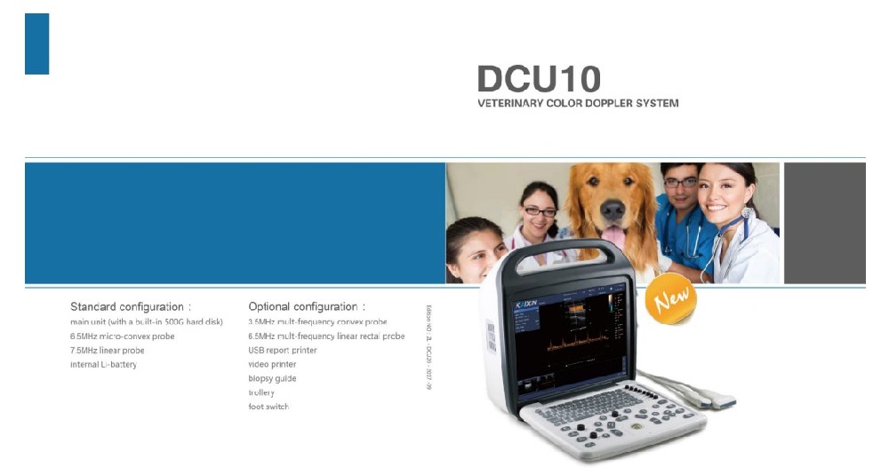
l 3.5MHz common abdominal probe: 1PC
l You can choose any 1pc probe from below :
>6.5MHz rectal linear probe
> 7.5MHz linear probe
>6.5MHz micro-convex probe
l Work station software : 1 Set
l Reticle:1 pc
12. Optional accessories
l Foot switch
l Portable trolley
l Video recorder
l Ethernet switch
If you have any questions, please contact us!
CONTACT US